
Microscope-Assisted Root Canal Treatments
Micro-endodontics or microsurgical endodontics is an aspect of the root canal procedure that evolved after the introduction of the Dental Operating Microscope (DOM) to endodontics in the late 1990s. One may ask how a microscope could assist a dental practitioner in performing a root canal?
The goal of root canal treatment
Severe tooth decay or other trauma create open gates for bacteria to reach the inner part of the tooth. When nerves and blood vessels in the tooth are damaged by bacterial contamination, the tooth, in most instances, becomes painful. The goal of root canal procedures (endodontic treatments) is to save a painful tooth by removing pulp tissue comprised of nerves and blood vessels from canals inside the tooth. The canals are then cleaned, shaped and filled with a special material, and the tooth is restored, usually with a crown. In some cases, the root tip must be removed surgically.
Easier said than done, as in most teeth, a canal containing nerves and blood vessels runs from the center of the crown of the tooth (the part you can see above the gumline) through the root and out into the jaw. Near the tip of the root, the canal branches into many smaller canals, similar to a river delta. All of these smaller branches of the canal must be completely cleaned and sealed in order to stop the pain and ensure a good and long-lasting outcome. These minute branches can be seen much easier under the microscope.
Benefits of micro-endodontics
After initially using the microscope in surgery, many endodontists (root canal specialists) discovered that it could also help with diagnosis and non-surgical endodontic procedures. Dental practitioners have used the microscope to find tiny painful fractures, which are often difficult to detect using traditional diagnostic methods. In addition, canals that are hidden, extremely very narrow or unusually positioned become easier to see. Occasionally, new infection or injury will cause a previously treated tooth to require a second endodontic procedure. Dentists call this “retreating” the tooth. To accomplish retreatment, all previous filling material and posts that may have been placed to support a crown must be removed from the tooth. The microscope may help with these procedures as well.
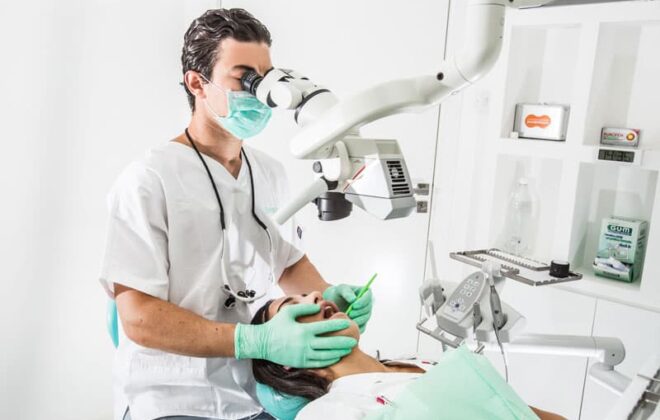
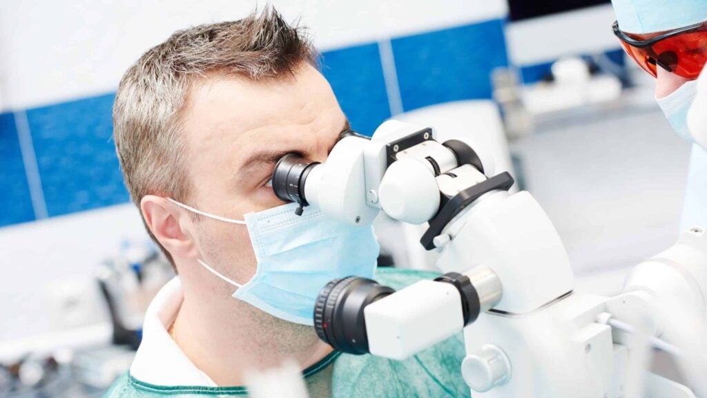
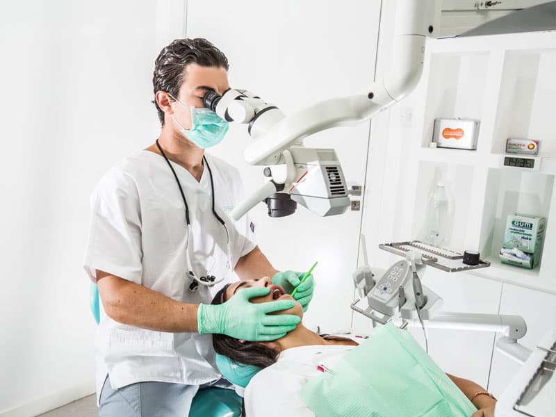
Why a dental operative microscope?
The recent addition of the dental operative microscope (DOM) to endodontic therapy allows for better visualization and management of the intricate morphology of the root canal system during endodontic procedures through magnification and greatly improved high-intensity lighting. The typical magnification range of the dental microscope is 4 to 25 times. The other commonly-used magnification aid, the surgical telescope (a tiny microscope mounted inside an eyeglass lens), magnifies between 2.5 and 4.5 times.
Surgical operating microscopes have a steep learning curve, and mastering them requires training as well as patience and practice. Still, this piece of equipment and the learning effort it implies are well worth it, as cases that once seemed impossible can now be treated with a high degree of confidence and clinical success.
Article originally appeared at: https://www.montreal-dentists.com/
Author: Montreal Dentists

